
Glaucoma
Glaucoma is the name given to a group of conditions whereby the optic nerve carrying the messages of sight from the eye to the brain becomes damaged and over time degenerates.
This degeneration is closely associated with an increase in the intraocular pressure (IOP), the cause of this is probably multi-factorial but is usually due to either an overproduction of aqueous humor or a slow drainage mechanism for the aqueous.
If you have been diagnosed with any form of AMD or feel like you may be developing symptoms, please get in touch with our team at Low Vision Centre by calling 01253 362696.
Types of Glaucoma
There are several different types of glaucoma, the main being:
- Primary Open Angle Glaucoma (POAG) – asymptomatic
- Acute Angle Closure Glaucoma (AACG) – very painful
- Latrogenic (caused by surgical intervention)
- Secondary to another disease
- Congenital or Developmental
This information page will concentrate on POAG and AACG in this list as these are the two forms which represent the vast majority of cases found in the UK.
Glaucoma is a major cause of sight loss in the UK and around the world, responsible for about 12% of all blind registrations in the UK. This is an unfortunate statistic because the majority of cases can be easily treated with simple eye drops, all it takes is a short eye pressure test to rule out the disease. If undetected it often causes significant sight loss before the patient is aware of any problems, so frequent eye examinations at the Optometrist are vital in the battle against this disease.
About 2% of people over the age of 40 suffer with glaucoma in one form or another and this increases to 10% of the population in the UK over 70 years of age. Family history is an aggravating factor as those with a parent suffering with glaucoma have a increase in risk of 4% and those with a sibling have a 10% increase in risk. Currently the NHS in England covers the cost of an annual eye examination for those with an immediate family member suffering with the disease in an effort to catch the disease development as soon as possible.
Different racial groups are also at higher risk of developing the disease with Afro-Caribbean heritage being the most at risk group. Those who have a hyperopic prescription (long-sighted) are also more susceptible. This is due to the eye being smaller, therefore, having less room to accommodate all the internal ocular structures resulting in a more compact eye and subsequently more risk of an increase in pressure with small changes in Aqueous production and or drainage.
Aqueous humor is a nutrient rich viscous fluid produced by the ciliary body which is situated just behind the iris. The role of the aqueous is primarily to serve the avascular tissues, ie the cornea, with nutrients such as amino acids.
Due to it’s continuous production it also has an affect in the intra-ocular pressure. After it’s production the aqueous flows between the iris and the intra-ocular lens, through the pupil, before circulating in the anterior chamber between the iris and the cornea. This is where it imparts it’s nutrients before draining away through the crevice formed between the cornea and the iris root.
This area contains the trabecular meshwork which is similar in structure to a microscopic sieve draining the fluid out of the eye. Should the meshwork become clogged or should the ciliary body over-produce the aqueous then of course a pressure will build up as the fluid is being made quicker than it can be drained away.

Intra-Ocular Pressure (IOP)
Manipulating the Intra-ocular pressure by medication and or surgical intervention is the method of treatment of all glaucoma.
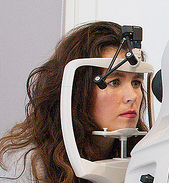
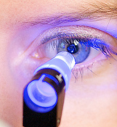
It is, therefore, important to understand what is a normal IOP. Some would argue that this is a whole degree subject in itself, however, for all intents and purposes a “normal” IOP should be between 6 and 21mmHg, any higher or lower could mean there is a problem with your eyes which needs investigating further. The pressure is taken using either an NCT (Non-Contact tonometer – puff of air test) or an Applanation Tonometer which is more accurate but more time consuming as anaesthetic eye drops are needed due to a small probe gently tapping the surface of the eye. We would only perform the second method if we were concerned about the condition of a persons eyes.
The pressure of the eyes is very variable and can even be influenced by the testing procedure itself. A person who is quite anxious about the puff of air test can give artificially high readings due to the squeezing action of the eyelids, this is known as white coat syndrome.
More commonly, the pressure can be affected by the following:
- Diurnal variation – higher in the morning due to natural physiology of the eye
- Fluid intake – drinking lots of water can have a temporary increasing affect on IOP
- Heart rate / Blood Pressure – can raise the IOP if themselves are high
- Alcohol – excessive intake can have a temporary decreasing effect
- Caffeine – excessive intake can have an increasing effect
- Exercise – some studies have shown that meditation can have a reducing effect but other sports can have raise the IOP temporarily
- Music – studies have shown that people who play woodwind/brass instruments can have a higher IOP possibly due to the raised sinus pressure from playing the instruments
- There has even been a study to suggest that business men who wear tight ties can have an increased IOP
All this serves to demonstrate how complicated the ocular system is. Just because the pressure of your eye is high or low at a particular time does not definitively mean that you have a problem.
Raised IOP does not have any symptoms until it reaches the 30mmHg mark or higher (see ACCG below), therefore patients suffering with Glaucoma are often completely unaware of it’s presence. For that reason we cannot stress enough the importance of regular eye examinations to monitor this fluctuation, particularly if you have a family history of the disease.
Eye pressure is not the only factor to consider as there are people who exhibit what is thought to be a normal eye pressure and yet still show optic nerve degeneration in line with Glaucoma, this is termed Low Tension Glaucoma. Vice-versa there are also people who exhibit a high IOP but show no evidence of optic nerve degeneration, this condition is termed Ocular Hypertension (OHT) and needs to be monitored carefully as it can develop into glaucoma over time.
Field of Vision test
The field of vision test is very important in both detecting the presence of glaucoma, ie if any damage to the nerve and therefore, the visual field, has occurred and also in monitoring the disease to ensure any treatment is successful.
The visual field is the area with which you view the world. Although we use the central area of our visual field to focus on things and view detail there is a significant area which we can see “out of the corner of our eyes” but has less detailed information for us.
It is the peripheral visual field that is affected first in glaucoma. Due to the structure of the optic nerve the damage occurs in the peripheral visual field first and due to it’s less detailed vision we cannot discern these subtle changes ourselves. It takes a special test called the visual field test to pick up any field loss caused by damage to the optic nerve.
The visual field test uses a machine that presents different coloured lights or patterns before the eyes and requires a response by the subject to affirm that they can see this stimulus. The machine then varies the position and brightness of the stimulus to discern the sensitivity of the retina in different areas.
The field of vision test is very important in both detecting the presence of glaucoma, ie if any damage to the nerve and therefore, the visual field, has occurred and also in monitoring the disease to ensure any treatment is successful.

The greyscales represent how bright the stimulus has to be for the subject to see it (white means the light is very dim i.e., good eye sensitivity, and black meaning the eye cannot detect light in that area). The grey/black dot on the right is the natural blind spot of the eye and the sensitivity of the eye naturally diminishes further out from the centre (the greyness increases peripherally) The image on the right is that of a typical Glaucoma field plot where the eye has lost sensitivity above the mid line of the line of sight.
The black dot on the left is the eyes natural blind spot but the dark areas above and to the right are parts of the retina that cannot signal to the brain their messages due to optic nerve loss or damage.

Primary Open Angle Glaucoma (POAG)

Primary open angle Glaucoma accounts for over 90% of the cases of Glaucoma seen in practice. It is defined as a raised pressure within the eye usually caused by a slow clogging of the drainage mechanisms in the anterior chamber.
There are no symptoms and once diagnosed it is a chronic life long condition and is usually treated using eye drops although in more unstable cases surgical options are available. These include using a laser to create a small hole in the iris designed to relieve the pressure of fluid flowing through the pupil and inserting an artificial drainage tube into the white part of the eye.
Regular monitoring is essential in any Glaucoma cases, and for these in the immediate family also.
Acute Angle Closure Glaucoma (ACCG)
Acute angle closure Glaucoma differs from POAG as it is a rapid onset and very painful condition, it is very unlikely to affect both eyes at the same time. The pain is such that it can cause nausea and the eye is usually very sensitive to light so much so that patients often feel the need to wear dark glasses or close the eye affected. Patients sometimes describe seeing halos around lights also. The eye will be very red especially around the edge of the iris and the cornea can look cloudy.
In ACCG the angle between the cornea and the iris collapses completely blocking off the drainage mechanisms of the eye. This then leads to a rapid build up of pressure which can cause swelling and acute pain. In most cases the pressure rises to over 30mmHg and is often over 50mmHg (normal being under 21mmHg).
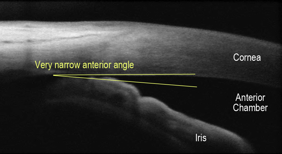
This condition needs URGENT medical attention to reduce the pressure and open the angle, usually using a laser to punch a hole in the iris.
Due to the high pressures involved if left for more than a few hours the optic nerve can be significantly and irreversibly damaged leading to visual impairment.
Anecdotally the condition can be caused by a large dilation of the iris as it crumples up much like a closed curtain. In a narrow angle this is enough to completely block the angle causing a rapid rise in IOP. This is often whilst the patient has spent time in a dark environment. It has been known that people suffer from this condition, if they are predisposed to it by having a narrow anterior chamber angle, after visiting the cinema or the theatre.
If you are concerned, please contact the practice so that one of our Optometrists can advise you of the best course of action. Alternatively, feel free to get in touch with our team at Low Vision Centre by calling 01253 362696.
LATEST NEWS EVENTS FUNDRAISING
Technology Exhibition 2026
N-Vision Assistive Technology Exhibition 2026 Discover the latest technology and support for living independently with sight loss N-Vision is inviting people living with sight loss,
Happy Retirement, Keith!
Celebrating Keith’s Retirement After 24 Years! Friday 21st August was a very special day at N-Vision as residents, staff, friends and family came together to
Technology with Confidence
Discover the Possibilities of Smart Glasses at N-Vision Special Technology Session for N-Vision Clients Technology is constantly changing, and new developments are creating exciting opportunities
